
Name horn or skin before you spray
Digital dermatitis and a sole ulcer fail entirely different tissues. One protocol cannot fix both.
3 min read · 2026-09-07
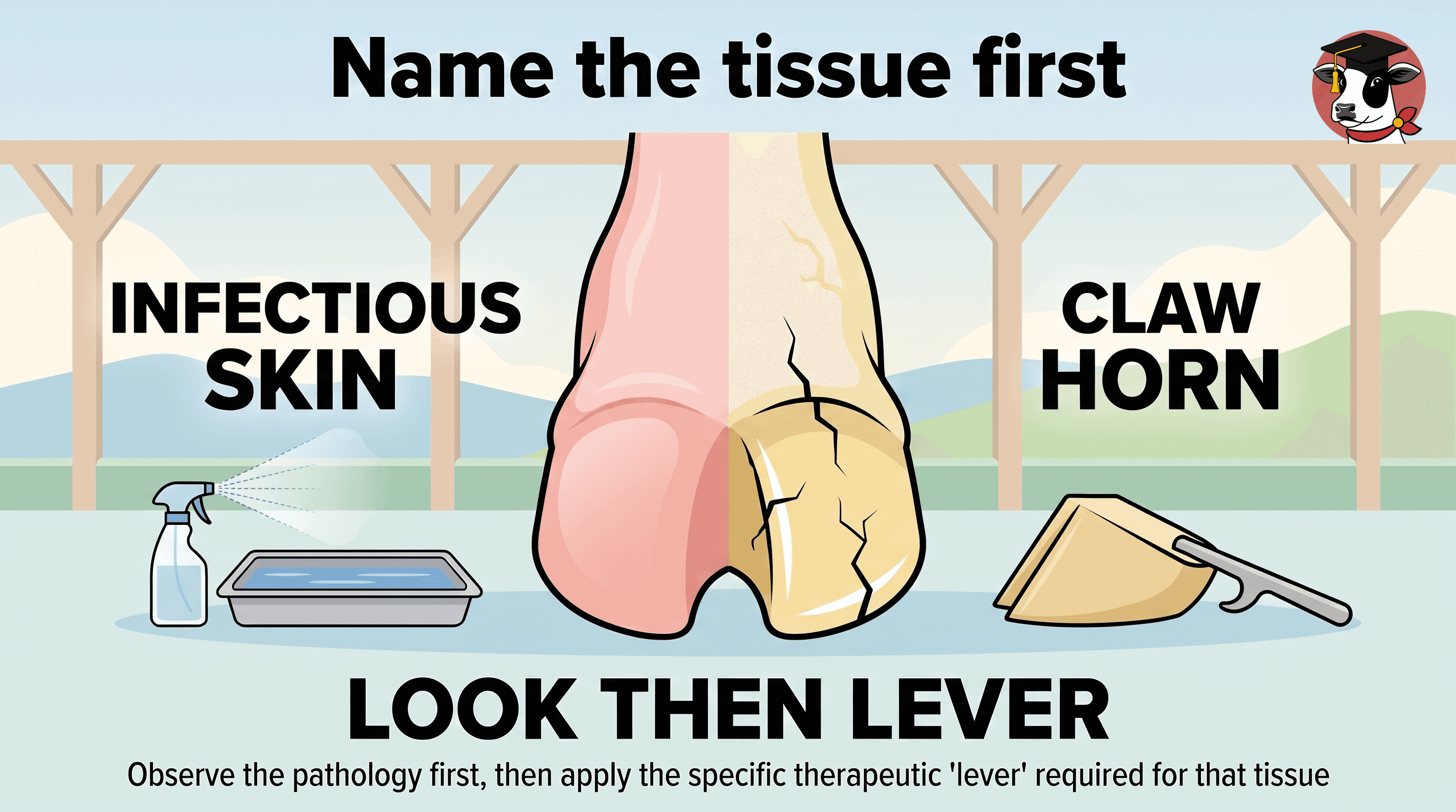
A common trap is treating every lame cow in the lockup with topical sprays, as if every lesion were infectious hairy heel wart. Literature separates infectious skin conditions (digital dermatitis, foot rot) from non-infectious claw-horn disruption lesions (sole ulcers, sole hemorrhages, white line disease).
The split is useful for first triage but not absolute. An ulcer begins as structural failure, yet exposed tissue in a non-healing lesion can become associated with Treponema. Complicated horn lesions may need both mechanical and infectious-disease assessment.
Topicals and well-managed footbaths fit surface-level infectious skin disease. Hygiene and footbaths can reduce digital dermatitis pressure. Active painful skin lesions need individual topical care under the herd veterinarian’s protocol. Chemicals do not repair damaged horn — spraying a sole ulcer treats mechanical failure as surface infection.
Claw-horn lesions reflect physical failure influenced by environment and metabolism. Transition BCS loss may reduce digital-cushion protection and raise sole-ulcer risk. Standing time is another lever: poor flow, abrasive flooring, long parlor holds, uncomfortable stalls. Protect lying time; review lockup and parlor routines.
Wash and inspect before classifying. Watch transition BCS, lying opportunity, flooring, lockup time. Infectious skin → topical/footbath pathways; claw-horn → professional trimming, offloading, pain planning with the veterinarian. Escalate non-healing lesions.
Sister notes on this universe cover the parlor that added two hours of standing, standing time as the dose, hoof records as herd trends, and camera scores as screening. Educational only — not a diagnosis, prescription, or treatment protocol.
Keep reading
View the networkNext
Hoof Blocks and Off-Loading: Taking Pressure Off the Painful Claw
A hoof block is mechanical off-load for a painful claw-horn lesion — not a spray substitute. Sound-claw selection, placement, and a timed recheck decide whether it helps or harms.
Sister note · 3 min
Home plate: Hoof · From lameness to lesion · Classify the lesion
- Classify the lesionHorn or skinPlaced here
- Camera scores are screening — not diagnosesComputer-vision gait scores can prioritize cows and track herd trends — useful screening when camera setup is honest, never a veterinary diagnosis on their own.Sister note
- Hoof records are herd trends — not diagnosesStandardized digital trim records and lesion codes can show infectious vs claw-horn patterns over time — useful herd trends when read with BCS, energy balance, and standing time, not a cow-side diagnosis.Sister note
Mark size is the kind. Color is the atlas area.
- Focus
- Field note
- Hoof
Sources
- Overview of Lameness in Cattle — Merck Veterinary Manual
- Digital Dermatitis, Sole Ulcers, and White Line Disease in Dairy Cattle: Pathogens, Diagnosis, and Treatment — Utah State University Extension
- Effects of routine treatment with nonsteroidal anti-inflammatory drugs at calving and when lame on the future probability of lameness and culling in dairy cows — Wilson et al., Journal of Dairy Science (2022)
- Review of White Line Disorders in Zone 3 and Toe Tip Necrosis in Dairy Cows and Recent Insights into Aetiopathogenesis and Treatments — Holzhauer & de Leeuw, Microorganisms (2025)
- “Non-healing” claw horn lesions in dairy cows: Clinical, histopathological and molecular biological characterization of four cases — Alsaaod et al., Frontiers in Veterinary Science (2022)