
Digital Cushion Thickness, Transition, and Sole-Ulcer Risk
The digital cushion helps dissipate load beneath the pedal bone. Thin sole soft tissues have been associated with later claw-horn lesions, but cushion thickness is not simply a reflection of body condition. Stage of lactation, parity, farm conditions, previous lesions, and tissue structure all appear to matter.
Knowledge level in this topic: Advanced. Mechanism and stacked risk in this topic — assumes the first-pass frame.
4 min read · 2026-09-17
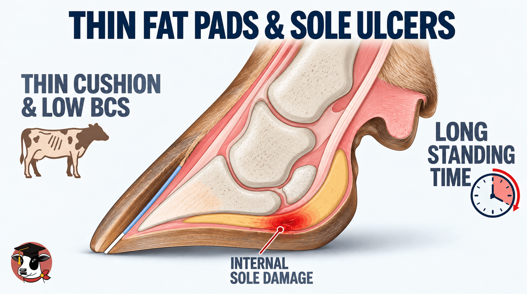
Sole ulcers are claw-horn lesions. Beneath the distal phalanx sits the digital cushion: cylinders of fat inside a collagen-rich connective-tissue framework. Together with the corium and the suspensory apparatus of the claw, it helps dissipate load before force reaches the horn-producing tissues. When that system is less able to spread load, the same standing day becomes a heavier mechanical challenge to the corium. Leave traction and wear to the flooring note, and hours on concrete to the standing-time note.
Ultrasound studies often measure the digital cushion together with the adjacent corium as sole soft-tissue thickness. A 2017 ultrasound study found that cows that experienced a sole ulcer or severe sole hemorrhage had sole soft tissues about 0.24 mm thinner than other cows, except when a sole ulcer was already present on that claw, when the tissues were thickened by about 0.53 mm — consistent with local inflammation. A follow-up analysis found thinner sole soft tissue ahead of a later sole ulcer or severe hemorrhage. A thin measurement can come before the lesion. A thicker reading after an ulcer appears does not mean the cushion is healthier.
Earlier studies linked greater body fat with thicker digital cushions, but newer prospective evidence shows that body condition alone does not reliably predict cushion thickness. Parity, stage of lactation, farm environment, lesion history, and tissue structure also matter. A 2009 study reported the body-fat association. The 2017 ultrasound study found sole soft tissues often thinner around calving than in late pregnancy, with a smaller backfat association that lesions could obscure. A 2024 prospective cohort of 2,352 cows on four UK farms found neither body condition score nor backfat thickness significantly associated with digital cushion thickness. Heifers ran thinner than multiparous cows until late lactation, and the thinnest point fell just after calving on some farms and in early lactation on others. A cow losing excessive condition through transition still deserves attention, because condition loss tracks lameness risk and wider transition trouble. The cushion does not simply thin whenever body fat is mobilized. Do not treat body condition as a proxy for cushion thickness.
The cushion is cylinders of fat embedded in a collagen framework, not a simple fat reservoir. A 2025 study found a lower proportion of type I collagen relative to type III in the digital cushions of cows with a history of lameness and hoof-horn lesions, and proposed that inflammation and remodeling can leave the tissue different after the original lesion has passed. Holding time, overcrowding, poor stalls, lock-up, slow parlor return, heat, and long walks all extend load under the pedal bone. A vulnerable cushion plus a long standing day is a different risk from the same cushion in a cow that gets enough lying time. The field lesson is that similar thickness is not the same mechanical protection.
Check in the barn
What to check: Are sole ulcers and severe sole hemorrhage concentrated in particular parities or stages of lactation? How much forced standing sits in the holding area, lock-up, treatment pen, and return alley? Are fresh cows using stalls, or standing in them? Do claw-horn lesions recur in the same cows across lactations? What does body-condition change say about transition management, without treating the score as cushion thickness? Are hoof records detailed enough to separate sole ulcer, hemorrhage, white line, and infectious disease? When a lesion appears, are you looking back at loading and transition history rather than only at today's ration?
Keep reading
View the networkNext
Sole Ulcers, Laminitis, and Diet: Separating Evidence from Assumption
Sole ulcers get blamed on “too much grain” more often than the evidence supports — separate diet risk from flooring, standing time, and claw mechanics before rewriting the ration.
Knowledge level in this topic: Advanced. Mechanism and stacked risk in this topic — assumes the first-pass frame.Sister note · 3 min
Home plate: Hoof · Why hoof lesions develop · Transition, body condition & the digital cushion
- Transition, body condition & the digital cushionThe pad changesPlaced here
- Flooring, Traction, Wear, and Their Effect on Claw HealthFlooring is a daily mechanical dose: traction and abrasive wear shape claw horn faster than most herds measure, and the wrong surface quietly builds the next ulcer cohort.Sister note
- Name horn or skin before you sprayDigital dermatitis and a sole ulcer fail entirely different tissues. One protocol cannot fix both.Sister note
Mark size is the kind. Color is the atlas area.
- Focus
- Field note
- Hoof
Sources
- Lameness in dairy cattle: A debilitating disease or a disease of debilitated cattle? A cross-sectional study of lameness prevalence and thickness of the digital cushion — Bicalho RC, Machado VS, Caixeta LS, Journal of Dairy Science (2009);92(7):3175–3184
- A prospective cohort study of digital cushion and corium thickness. Part 1: Associations with body condition, lesion incidence, and proximity to calving — Newsome RF, et al., Journal of Dairy Science (2017);100(6):4745–4758
- A prospective cohort study of digital cushion and corium thickness. Part 2: Does thinning of the digital cushion and corium lead to lameness and claw horn disruption lesions? — Newsome RF, et al., Journal of Dairy Science (2017);100(6):4759–4771
- A prospective cohort study of factors associated with the digital cushion thickness in dairy cattle — Griffiths BE, et al., Journal of Dairy Science (2024);107(9):7230–7244
- A history of lameness is associated with reduced proportions of collagen type I relative to type III in the digital cushions of dairy cattle — Wilson JP, et al., Journal of Dairy Science (2025);108(12):13807–13819